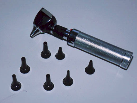
NB in textbooks and on this website, images of the ear are usually taken with an endoscope which has a wide angle lens on the end. This gives us an image which contains the whole tympanic membrane and much of the ear canal. When using an otoscope, you will NEVER get a view as good as this, not because it is your fault, but because of the optics of most endoscopes. It is unlikely that you will in fact be able to see the whole drum in one position. To see all aspects of the drum you will need to change position and move the tip of the otoscope.
Otoscopes
Ear drum shape
The Normal Eardrum
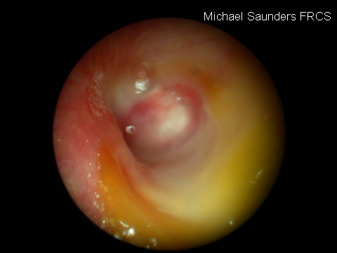
This diagram shows some of the features you can expect looking at a normal drum. There is no substitute for looking at as many drums as you can to give you an idea of what is normal and what is abnormal. Look in your fellow students ears to start with.
Shape of the Eardrum
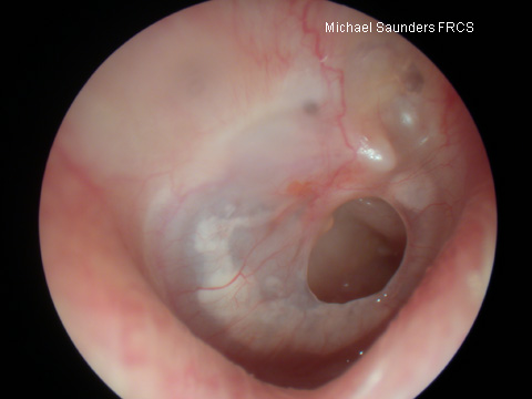
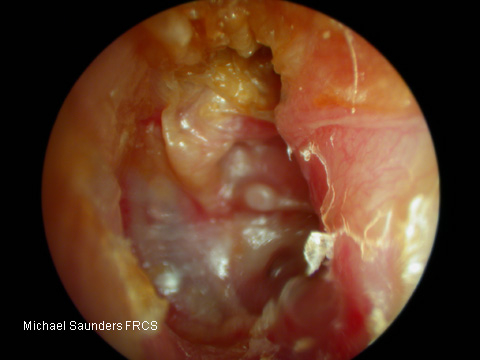
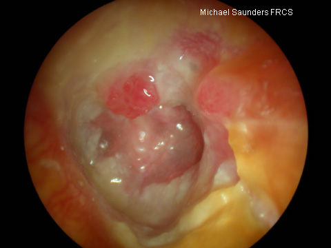
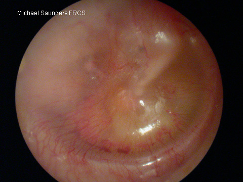
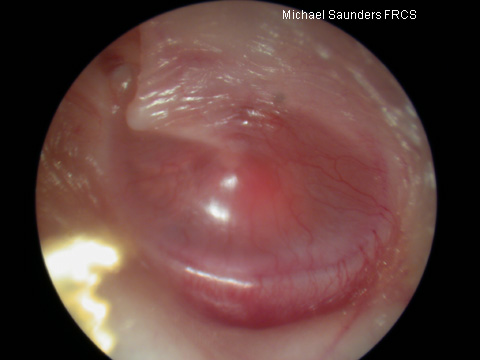
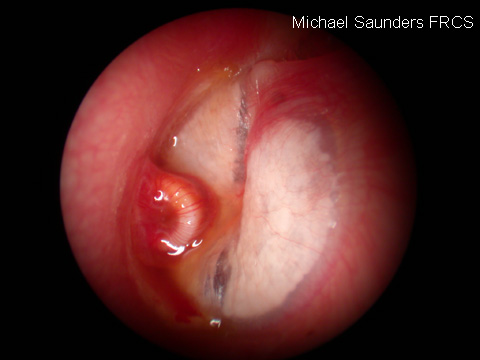
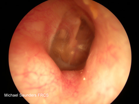
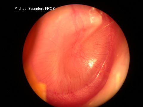
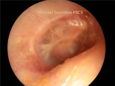
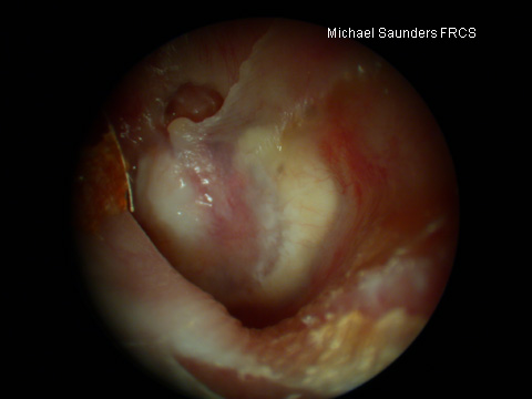
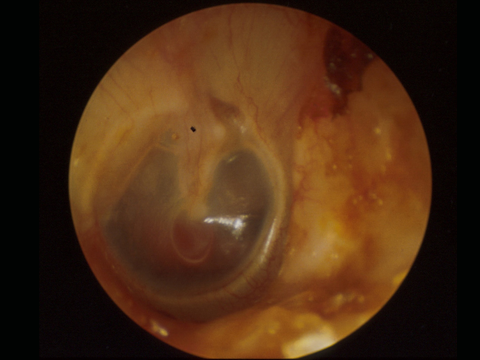
The drum is slightly convex being most medial at the end of the malleus handle or umbo. Abnormalities of shape are important, the drum may be bulging our, suggesting increased middle ear pressure, such as in acute otitis media, or retracted inwards with negative middle ear pressure, which is one of the otoscopic findings in glue ear.
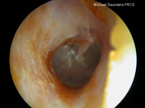
Bulging ear drums are usually fairly obvious and a result of increased middle ear pressure. The most likely cause of this is acute otitis media when the drum will not only bulge outwards, but is usually very red because of hyperaemia and infection. Unfortunately I have found good images of acute otitis media hard to come by because the patients are usually very young and not over disposed to sit still to have pictures taken!
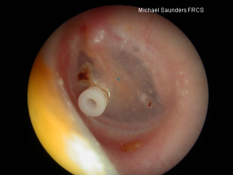
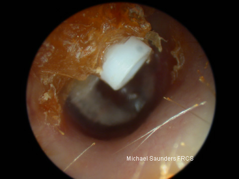
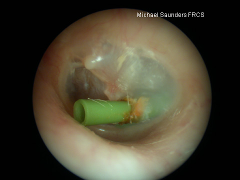
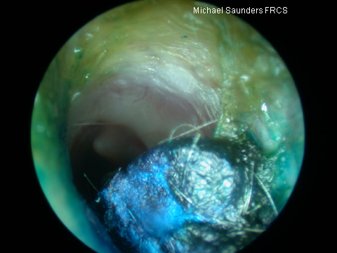
GROMMETS
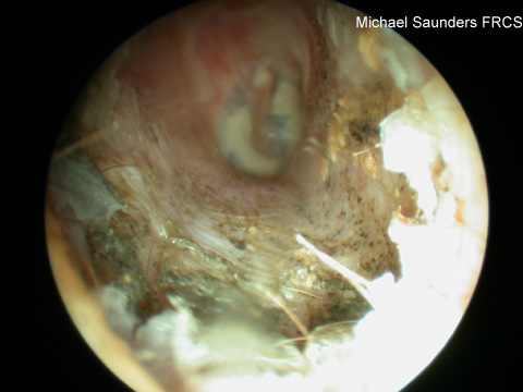
Ear canal
EAR DRUM COLOUR
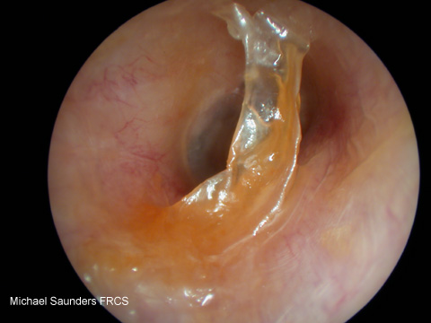
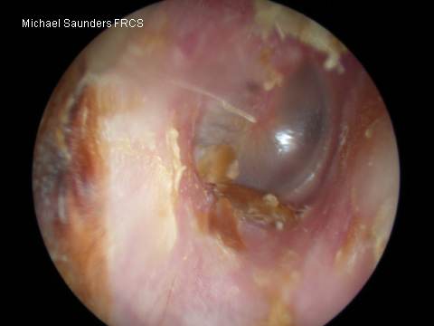
Colour of the Eardrum
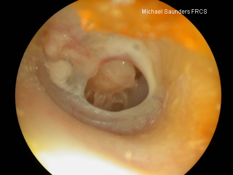
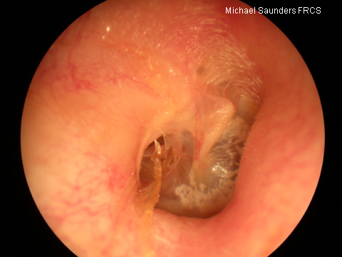
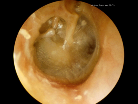
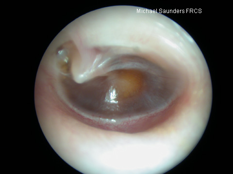
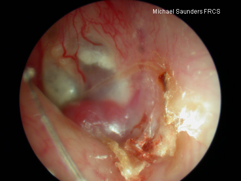
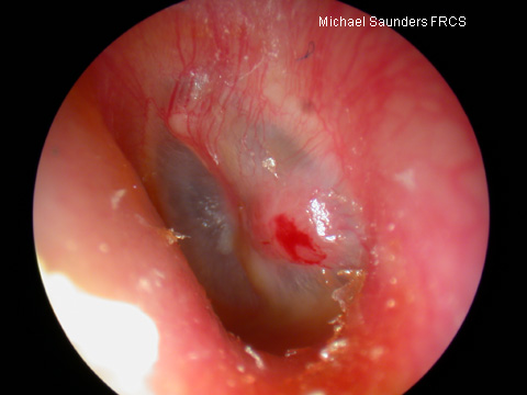
The normal drum is quite translucent and does not really appear to be any colour except perhaps grey. The colour of the drum can be changed by thickening of the drum itself, injection of blood vessels, or the presence of something behind it such as glue, pus or blood.

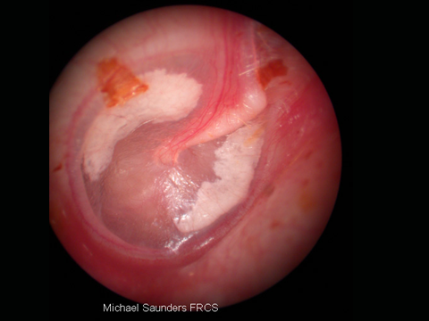
EARDRUM PERFORATIONS
You need to be able to distinguish between safe and unsafe perorations. A safe perforation is exactly what it sounds like: a hole in the tympanic membrane. The main risk of safe perforations are that they may allow infection to enter the middle ear but there are rarely more serious sequelae.